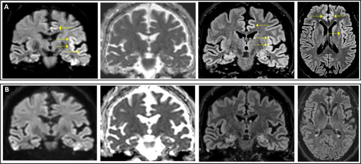
Figure 2.

Representative MRI images showing coronal DWI, ADC, and FLAIR sequences of the same slice and axial FLAIR sequence, performed in the subacute phase (day 10; (A) and post‐acute phase (day 50; (B). Abnormal cortical areas are indicated by arrows. Notably, ADC map does not show commensurate hypointensity in the anterior cingulate and insula, where the DWI and FLAIR cortical hyperintensity was present. ADC, apparent diffusion coefficient; DWI, diffusion‐weighted imaging; FLAIR, fluid‐attenuated inversion recovery.
