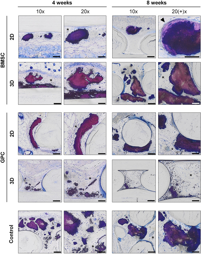
FIGURE 7.
Undecalcified histology. Representative images of BMSC, GPC and control constructs (no cells) after 4 and 8 weeks (thin ground sections, Levi Lazko staining); * indicate blood vessels; arrow indicates the only instance of “bone-like” tissue observed in the study; scale bars 100 μm (10x) and 50 μm (20x).

