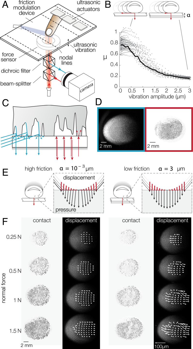
Fig. 1.
(A) Experimental setup. The friction between the fingertip and glass plate is reduced in the presence of flexural ultrasonic waves. A dual-illumination setup where blue light illuminates the skin at a 20 angle and red light is normally incident to the glass surface. (B) When a fingertip slides across the glass plate, the friction coefficient is reduced with increasing ultrasonic amplitude. The black line represents the median friction coefficient. (C) Close-up view of the illumination combining a dark-field blue light to highlight the fingerprint ridges and a red light, coaxially oriented with the camera, to illuminate only the asperities of the skin in intimate contact with the glass plate. (D) Typical images of the fingertip profile (Left) and the asperities in intimate contact (Right). (E) Presumed deformation of the skin when pressed against the surface in high- and low-friction conditions. Point trajectories are shown in red. The black arrows represent the pressure and traction exerted by each point on the surface. (F) Images of the intimate contact and skin deformation for increasing normal forces (top to bottom) and the highest (Left) and lowest (Right) friction. The white arrows show the displacement of reference points, scaled up 10-fold.

