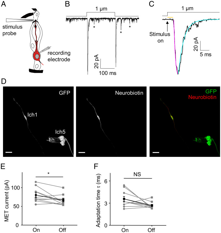
Fig. 1.
Current responses of Cho neurons to mechanical stimuli. (A) Schematic illustrating the patch clamp recording preparation of Cho neurons; the illustration of Cho neurons was adapted from ref. 2. Copyright [2000] Society for Neuroscience. A glass probe driven by a piezo actuator was used to deliver mechanical stimuli to the tip of the lch1 Cho neuron dendrite. The cell in red is the lch1 neuron. (B and C) The whole-cell current in lch1 neurons was evoked by a 1-μm stimulus. The asterisks mark the spontaneous discrete depolarizations. The “on” response current is shown in C at a higher time resolution. The arrow represents the onset of the stimulus. The orange line indicates the response latency of the MET current. The purple line represents a single exponential fit of the activation of MET currents; the cyan line represents a single exponential fit of the adaptation of MET currents. (D) Post hoc immunostaining of the targeted neuron loaded with neurobiotin. Iav-Gal4 was utilized to drive the expression of GFP in Cho neurons. (Scale bar, 15 μm.) (E and F) The amplitudes of the “on” and “off” current are shown in E (n = 10; paired t test). The peak values (absolute value) are used here. The adaptation time constants (τ) are summarized in F (n = 10; paired t test). All error bars denote ± SEM. *P < 0.05; NS, not statistically significant. The holding potential was −60 mV, and the MET current was recorded in extracellular Na+/intracellular K+-based solutions. Genotype is as follows: Iav-Gal4/+; UAS-CD8-GFP/+.

