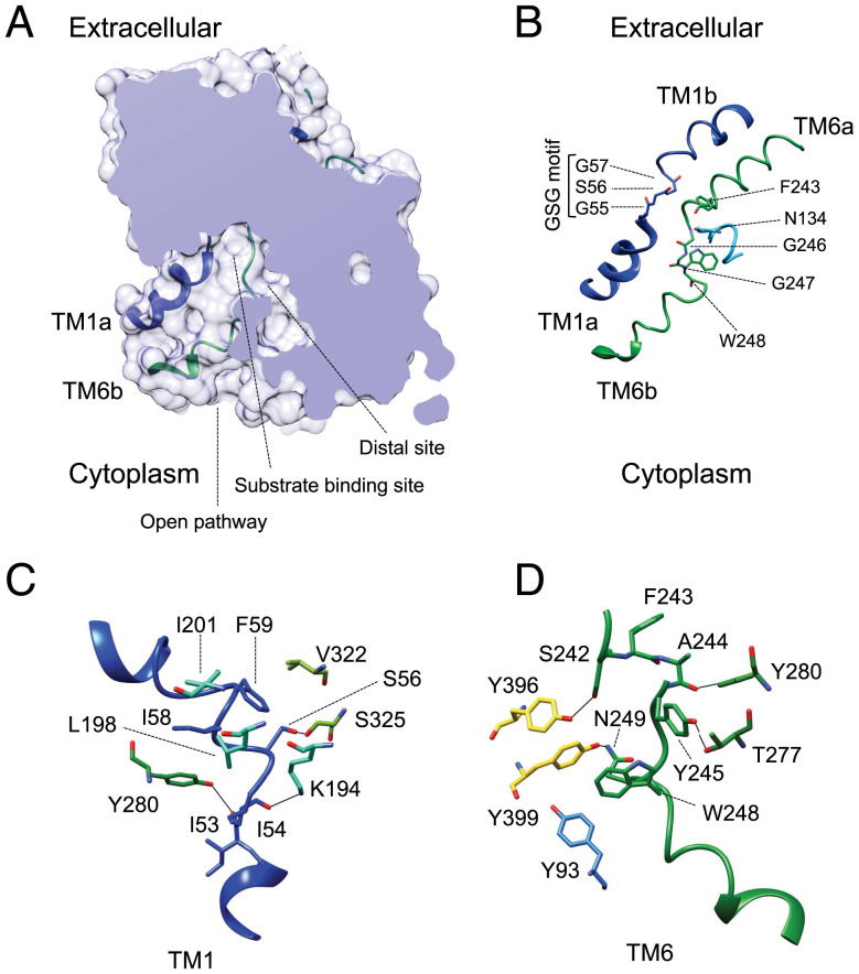
Fig. 2.
Unwound regions in TM1 and TM6 form the substrate-binding site opened to the cytosol. (A) Structure of hLAT2, represented as a surface, with a section to show the central cavity harboring the substrate-binding site opened to the cytoplasm but without access to the extracellular space. The TM1a and TM6b helical regions that open the vestibule are shown superimposed. A distal cavity connects to the central vestibule. (B) TM1 and TM6 forming the substrate-binding site are shown as a cartoon with key residues highlighted. The contribution of N134 from the adjacent helix TM3 to the substrate binding is shown. (C) The conformation of TM1 is maintained by interactions with residues in the vicinity. (D) Interactions between TM6 and neighboring regions of the structure. Color codes for hLAT2 helices and residues in all the panels are as used in Fig. 1. Oxygen atoms are shown in red and nitrogen atoms in blue.

