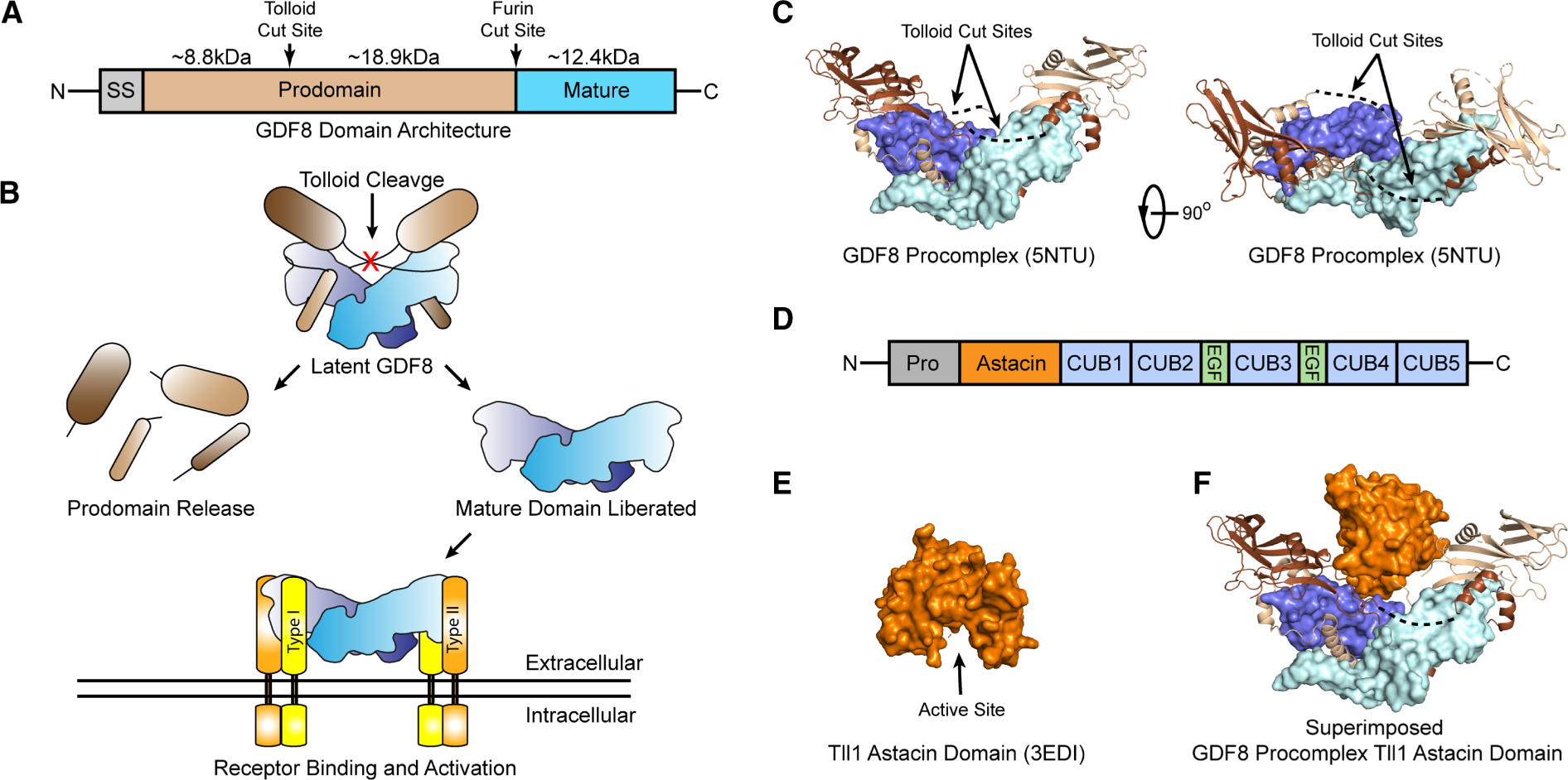
Figure 1. Latent GDF8 activation and Tll1 astacin domain structure.

(A) Domain architecture of GDF8, signal sequence (SS), prodomain and mature domain. Tolloid and furin cut sites indicated with size in kDa of each fragment after furin and tolloid processing shown. (B) Schematic of latent GDF8 activation, including tolloid cleavage, mature domain release, and receptor binding. (C) Structure of the GDF8 procomplex [27]. Mature domain monomers in pale cyan and blue, prodomain monomers in light brown and brown. Dotted lines indicated resides not in density. Right panel is rotated 90° about the vertical axis. (D) Domain architecture of the tolloid family including the prodomain, astacin domain, CUB and EGF domains. (E) Structure of the Tll1 astacin domain, active site cleft shown [58]. (F) GDF8 prodomain structure shown in (c) superimposed with the astacin domain active site cleft positioned toward the scissile bond.
