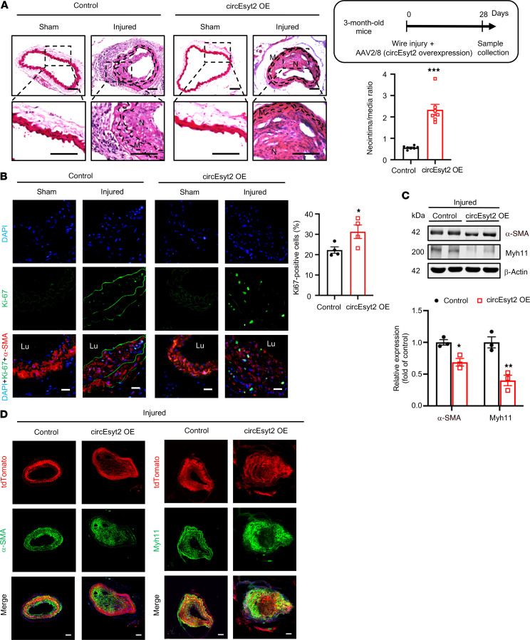
Figure 4. Effect of circEsyt2 overexpression on arterial remodeling in vivo.
(A) Wire injury was accompanied by injection of circEyst2-overexpressing (circEsyt2 OE) or control virus (control) in the carotid arteries from C57BL/6J mice. Carotid arteries were collected 28 days after injury. Left: representative H&E staining images. Scale bars: 50 μm. Right: ratio of neointima to media thickness of carotid arteries. ***P < 0.001 vs. control. n = 7. The insert on the right upper side depicts the schema of the experimental design. (B) Immunofluorescence staining for Ki67 in injured carotid arteries, treated as in A. Left: representative immunofluorescent images. Scale bars: 20 μm. Right: Ki67-positive cells in the injured group. *P < 0.05 vs. control. n = 4. (C) Western blotting to check for the expression of α-SMA and Myh11 in injured carotid arteries from C57BL/6J mice, treated as in A. *P < 0.05, **P < 0.01 vs. control. n = 3. (D) Wire injury was performed in circEsyt2-overexpressed carotid arteries with Tagln-CreERT2/tdTomato under the control of tamoxifen. Immunofluorescence staining for α-SMA or Myh11 in carotid arteries 28 days after injury. α-SMA or Myh11 (green), tdTomato (red), and DAPI (blue) were merged. Scale bars: 50 μm. Data are mean ± SEM. Two-sided unpaired t test for A and C. Kolmogorov-Smirnov test for B.

