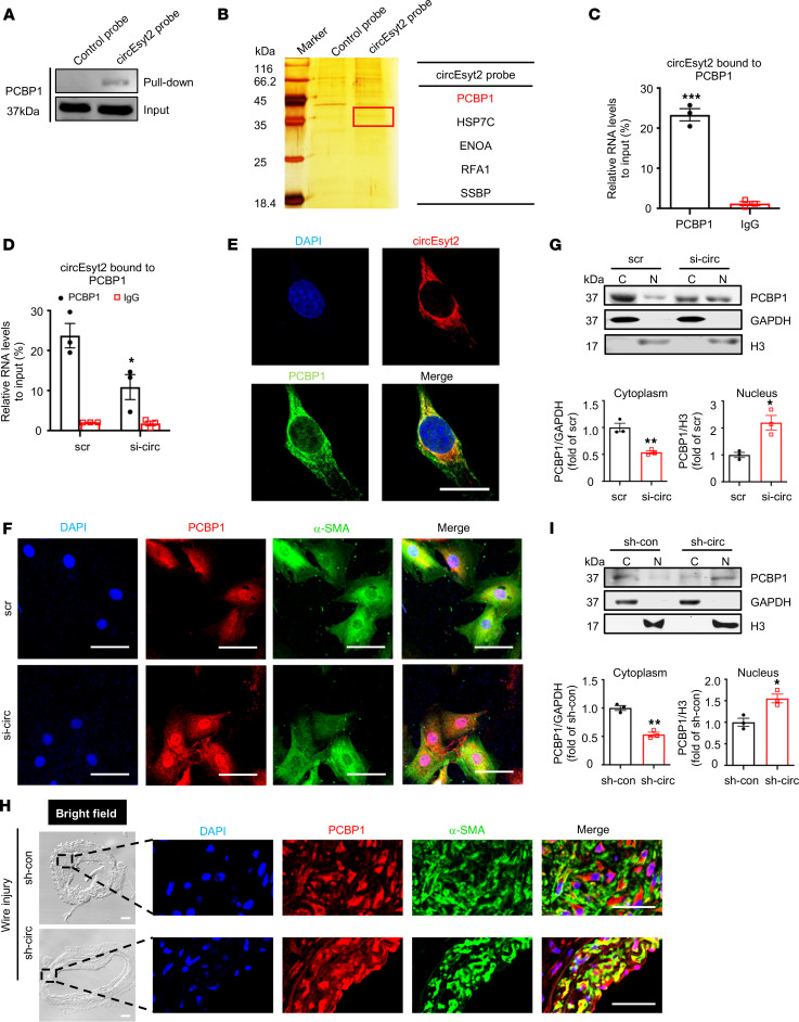
Figure 7. CircEsyt2 inhibits the nuclear trafficking of PCBP1 by binding directly to PCBP1.
(A) Western blotting of proteins pulled down by control and circEsyt2 probes in circEsyt2-OE HEK293T cells using the PCBP1 antibody. (B) Identification of circEsyt2-binding proteins. Left: silver staining of pulled-down proteins in circEsyt2-OE HEK293T cells. Right: mass spectrometry showing the main proteins pulled down by the circEsyt2 probe. (C) RIP-qPCR assay confirming the direct binding of PCBP1 to circEsyt2 in HASMCs. ***P < 0.001 vs. IgG. n = 3. (D) RIP-qPCR assay detecting the specific binding of PCBP1 and circEsyt2 in HASMCs by circEsyt2 silencing. Scrambled siRNA (scr) served as control. *P < 0.05 vs. scr. n = 3. (E) FISH of circEsyt2 (red), PCBP1 (green), and DAPI (blue) in HASMCs transfected with circEsyt2 plasmid. Scale bars: 20 μm. (F) Coimmunofluorescence of PCBP1 (red), α-SMA (green), and DAPI (blue) in circEsyt2-silenced HASMCs. Scale bars: 50 μm. (G) Western blotting to check the expression of cytoplasmic (C) and nuclear (N) PCBP1, treated as in F. Cytoplasmic control: GAPDH; nuclear control: histone 3 (H3). *P < 0.05, **P < 0.01 vs. scr. n = 3. (H) Coimmunofluorescence staining for PCBP1 (red), α-SMA (green), and DAPI (blue) in injured carotid arteries of the control (sh-con) and circEsyt2 knockdown (sh-circ) groups. Scale bars: 50 μm (bright field) and 20 μm (immunofluorescence). (I) Western blotting to check the expression of cytoplasmic and nuclear PCBP1 in carotid arteries, treated as in H. *P < 0.05, **P < 0.01 vs. sh-con. n = 3. Data are mean ± SEM. Two-sided unpaired t test for C, D, G, and I.

