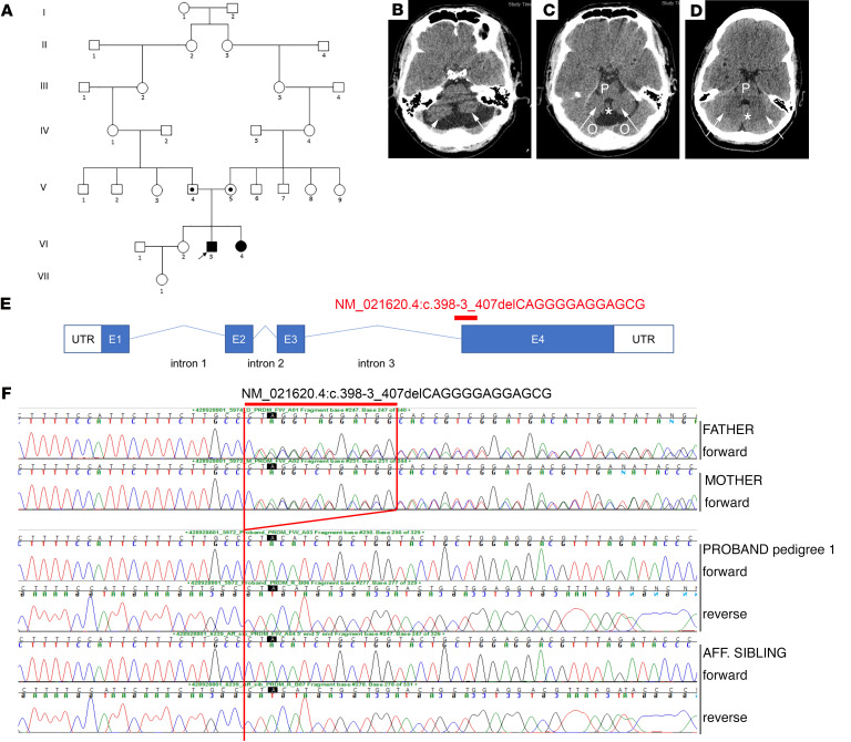
Figure 1. Exome sequencing identifies a PRDM13 mutation in 3 patients from 2 Maltese pedigrees.
(A) Pedigree 1 with 1 affected male (VI.3) (patient 1) and 1 affected female (VI.4) (patient 2) with a syndrome associated with HH and cerebellar hypoplasia carrying a homozygous PRDM13 mutation. Circles denote females; squares denote males; black square denotes affected male, and black circle denotes affected female; a dot in the middle of a shape indicates a heterozygous carrier; arrow indicates the proband. (B–D) (B and C) Axial slices on CT scan of the brain showing cerebellar hypoplasia in patient 1 compared with a normal CT scan from an unrelated individual (D). Arrows in B and C demonstrate hypoplastic cerebellar lobes as compared with those in the control scan (D). The pons (P) is hypoplastic compared with the scan in D, as is the cerebellar vermis (asterisk). Partial voluming from the occipital lobes above the tentorium is seen (O). (D) CT scan showing normal cerebellar lobes (arrows), a normal cerebellar vermis (asterisk), and a normal pons (P). (E) Diagram of PRDM13 transcript (NM_021620.4) showing the deletion found in patients at the intron 3/exon 4 border, which is predicted to affect splicing and to form a truncated PRDM13 protein. (F) Electropherograms of patients 1 and 2 and their unaffected parents (pedigree 1) showing the c.398-3_407delCAGGGGAGGAGCG deletion homozygous in the 2 patients and heterozygous in the healthy parents. Aff, affected.

