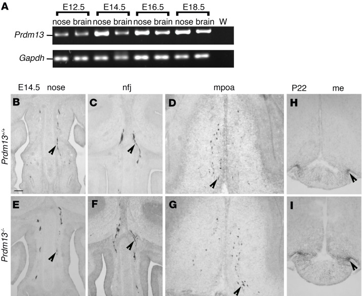
Figure 2. GnRH neuron specification and migration are not affected in Prdm13 mutant mice.
(A) RT-PCR analysis of Prdm13 expression in mouse embryonic nose and brain tissues extracted from indicated embryonic stages of WT mouse embryos. Gapdh expression serves as positive control. W, water-only control, no cDNA. (B–G) Coronal sections of Prdm13+/+ and Prdm13–/– E14.5 heads, immunolabeled for GnRH to reveal GnRH neurons in the nasal compartment (B and E), in the nfj (C and F), and in the mpoa (D and G). Black arrowheads indicate examples of neurons migrating in the nasal compartment (B–E), crossing the olfactory bulbs (C and F), and reaching the mpoa (D and G). (H and I) Coronal sections of Prdm13+/+ and Prdm13–/– P22 brains immunolabeled for GnRH to reveal median eminence (me) innervation by GnRH neuron neurites, indicated by black arrowheads. Scale bar: 250 μm.

