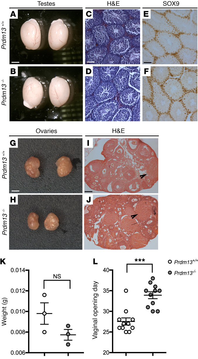
Figure 4. Prdm13-deficient mice display normal gonadal structure, but delayed vaginal opening.
(A and B) Microphotograph of testis pairs of the indicated genotypes; no differences were observed in their size. (C–F) H&E-stained (C and D) and SOX9-immunostained (E and F, Sertoli cells marker) testis representative sections from adult male mice of indicated genotypes. No differences and normal spermatogenesis were observed in WT and mutant mice. (G and H) Microphotograph of ovary pairs of the indicated genotypes. (I and J) H&E-stained ovary representative sections from mice of indicated genotypes. No differences in corpora lutea number were observed between WT and mutant mice. (K) Weight of ovaries from adult female mice. No differences were observed between WT and mutant mice (2-tailed unpaired Student’s t test). (L) Age at the time of the vaginal opening of the indicated genotypes. Note the significant delay in vaginal opening of Prdm13–/– female mice. ***P < 0.001, 2-tailed unpaired Student’s t test. Scale bars: 1.5 mm (A, B, G, and H); 250 μm (C–F); 500 μm (I and J).

