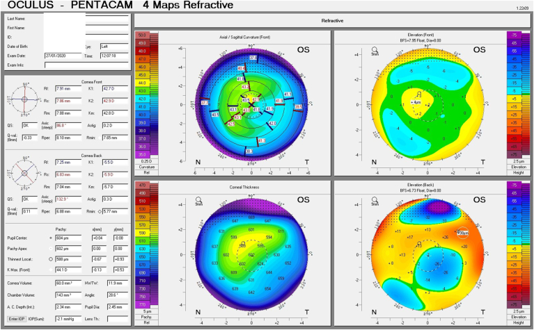
Fig. 2.
Tomographic maps before treatment exhibiting in the right eye focal posterior corneal surface depression, displacement of the thinnest point of the cornea, irregular isopachs in the right and left eye(A and B).Tomographic maps after eight days of treatment exhibiting in significant decrease in corneal thickness in the right and left eye(C and D).


