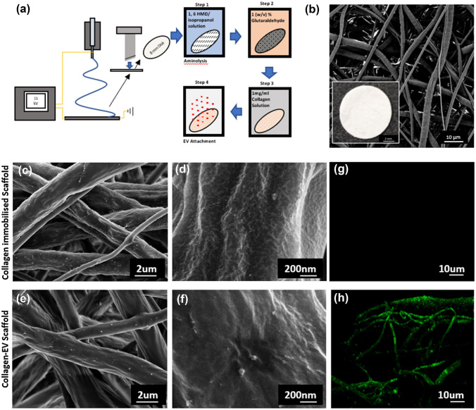
Figure 3.
Electrospun scaffold characterization, functionalization and EV attachment. (a) Schematic diagram of electrospinning and functionalization process. (b) SEM image of 8 mm electrospun PCL scaffold (inset: macro image of scaffold). (c–f) SEM imaging of scaffold fibre morphology of collagen functionalised and collagen-EV functionalised scaffolds respectively. (g and h) Confocal imaging of collagen-functionalized scaffold after incubation with a PKH-26 dye control or PKH-26 labelled EVs respectively.

