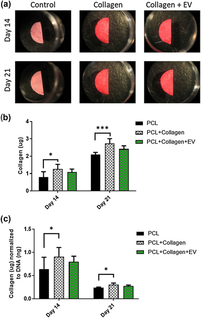
Figure 5.

The effects of PCL scaffold surface modifications on collagen extracellular matrix deposition. (a) Picosirius red staining of PCL, PCL + Collagen, and PCL + Collagen + EV scaffolds after 14- and 21 days culture. (b) Quantification of collagen matrix after 14 and 21 days. (c) Quantification of collagen synthesis normalised to DNA content after 14 and 21 days. All data n = 6. Statistical analysis using two-way analysis of variance (ANOVA) and Tukey's multiple comparison post‐test (*p < 0.05, ***p < 0.001). EVs extracellular vesicles.
