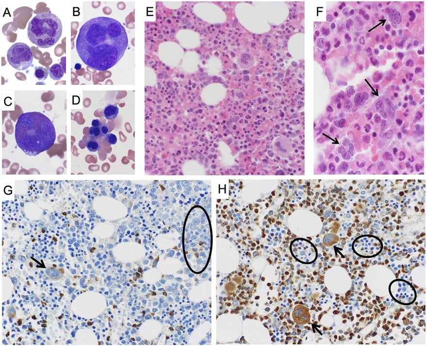
Figure 2: Hematopathological features SEPT6-associated congenital MDS and abnormal bone marrow SEPT6 staining corrected after allogenic HSCT.
Bone marrow aspirate and biopsy of the patient displayed strikingly abnormal myeloid precursors which were diffusely present. In BM aspirates stained by May-Grunwald-Giemsa (MGG, at 40x magnification), we observed giant multinucleated neutrophils (panel A), giant multinucleated promyelocytes (panel B), giant multinucleated eosinophils (panel C). In addition, we noted prominent nuclear lobation in erythroid precursors (panel D). In parallel, similar findings were seen on BM biopsy after hematoxylin & eosin (H&E), confirming the presence of abnormal, giant myeloid progenitors (panel E, 20x magnification) and multinucleated granulocyte precursors (panel F, 40x magnification, arrows). Bone marrow biopsies from the patient were stained after validation of a SEPT6 antibody for immunohistochemistry on an array of normal human tissues (Supplementary Figure S6). In the biopsies stained by MGG prior to HSCT (panel G, at 20x magnification), we observed markedly decreased SEPT6 staining in granulocyte precursors (circle) and megakaryocytes (arrow). This abnormality was corrected in the biopsy post-HSCT (panel H, at 20x magnification), where we noted normal SEPT6 staining of myeloid progenitors and megakaryocytes (arrows), and comparatively decreased erythroid staining (circles).

