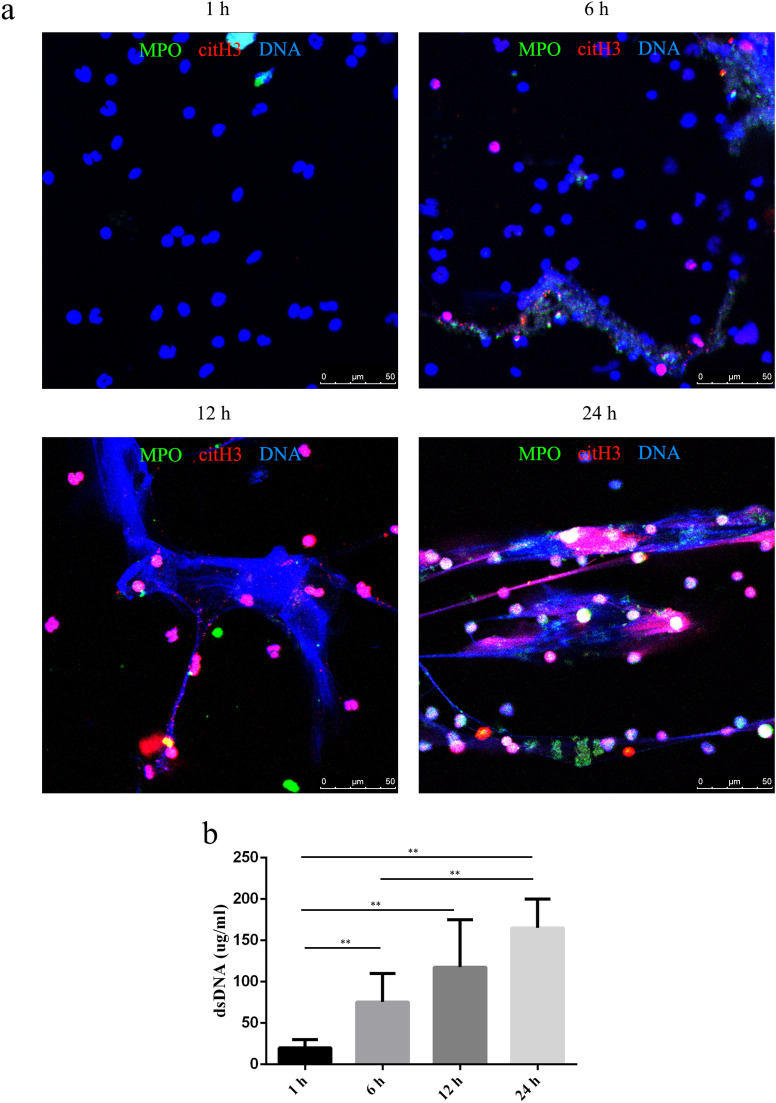
Figure 1.
Aging-related spontaneous NETosis. The proportion of spontaneous NETs and dsDNA released by neutrophils was evaluated by fluorescence microscopy (a) and PicoGreen staining (b) at 1, 6, 12, and 24 h. (a) Samples were stained with antibodies against citrullinated histone H3 (citH3, red) and myeloperoxidase (MPO, green) and DNA was counterstained with DAPI (blue). Original magnification, 40× ; scale bar, 50 μm. (b) DNA release was compared between different groups, and the data are represented as the median with range from six independent experiments, **P < 0.01.

