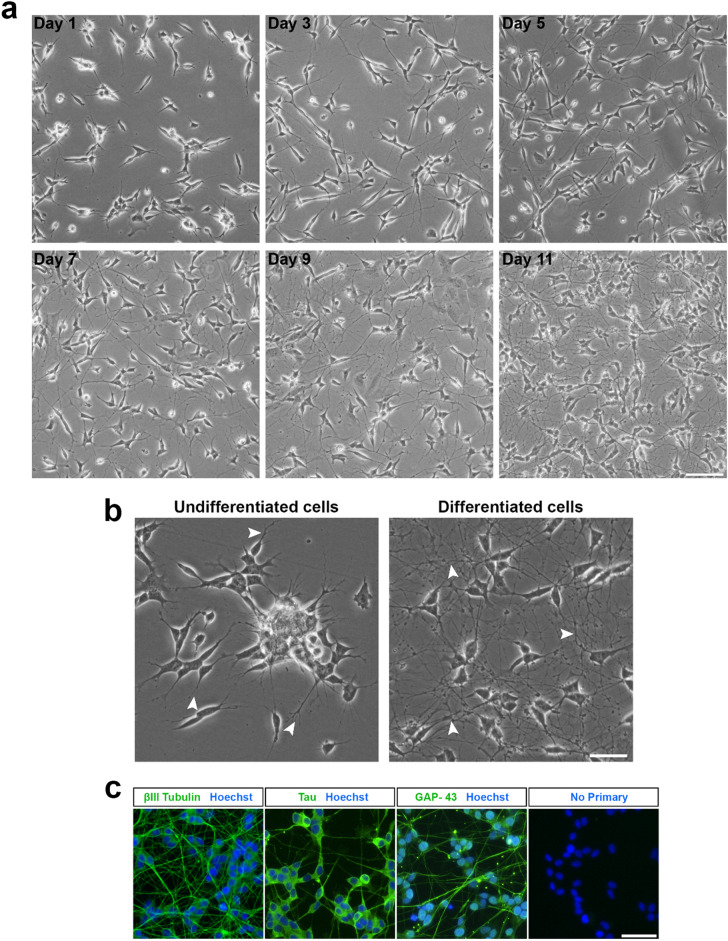
Figure 1.
Differentiation of the SH-SY5Y cell line into a neuronal phenotype. (a) Phase contrast images of SH-SY5Y cells at various stages of the differentiation process. Cells were treated with RA (10 µM) for five days, followed by BDNF (50 ng/mL) for an additional five days to achieve terminal differentiation by Day 11. Images were acquired using an EVOS FL microscope at ×10 magnification and 43% brightness (scale bar = 50 μm). (b) Undifferentiated SH-SY5Y cells possess few short projections and cluster together, while differentiated cells are observed to have many extensive projections. Images were acquired at ×20 magnification and 60% brightness using an EVOS FL microscope (scale bar = 25 μm). βIII-tubulin images were acquired using the GFP filter (excitation 482/25 nm; emission 524/24 nm; exposure 110 ms), while Hoechst nuclei images were acquired using the DAPI filter (excitation 357/44 nm; emission 447/60 nm; exposure 19 ms). White arrowheads point towards neurite extensions. (c) Expression of mature neuronal markers by differentiated cells. Fluorescence images were acquired at ×20 magnification using an EVOS FL Auto microscope (scale bar = 25 μm).

