Figure 3.
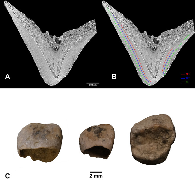
Virtual histology. (A) Vestibular-lingual section of the AVH-1g deciduous upper first molar passing through the mesio-lingual cusp (pixel size = 3 µm, reformatted slice thickness 30 µm); (B) The same section showing position of the neonatal line (NL in green) and two accentuated lines (AL1 & AL2 in red and blue, respectively). Position of the NL allowed estimation of AVH-1’s age at death based on enamel development. The two accentuated lines reflect prenatal stress events. (C) Three deciduous teeth investigated through synchrotron X-ray computed microtomography; from left to right: AVH-1d (upper central incisor, vestibular view), AVH-1c (upper lateral incisor, vestibular view), AVH-1g (upper first molar, occlusal view).

