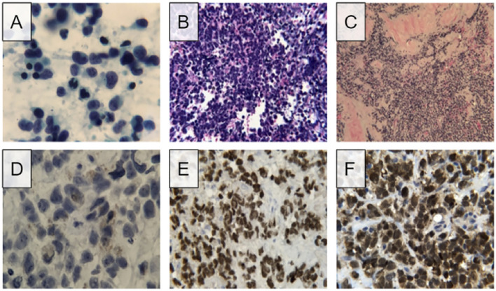
Figure 1.
Microscopic findings and immunohistochemistry stains of initial lymph node FNA and subsequent needle-core biopsy. (A) Direct smear of lymph node FNA shows pleomorphic cells with scant sytoplasm and fine chromatin, presence of nucleoli was not detected. (B) Routine hematoxylin and eosin staining of cell block visualizes numerous small cell populations with high nuclear to cytoplasmic ratio. (C) Routine hematoxylin and eosin staining of needle-core biopsy shows populations of small pleomorphic cells. (D) High magnification image of needle-core biopsy displays golden-brown coloration resembling that of melanin. (E) Positive result for SOX10 staining, a biomarker for melanoma. (F) Positive results for S100 staining, another biomarker for melanoma.

