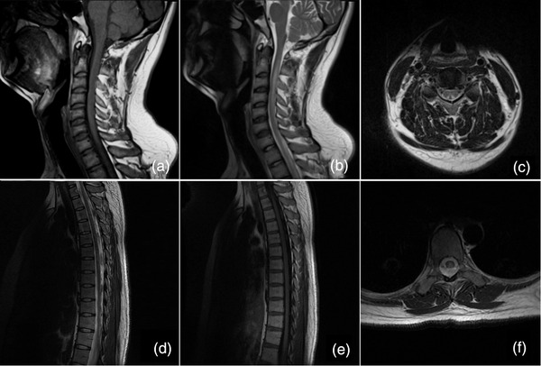
FIGURE 1.

Conventional magnetic resonance imaging of the spinal cord. A 24‐year‐old man presented with impairment but was able to walk unsupported and numbness in all extremities. Sagittal T1‐weighted imaging showed decreased intramedullary signal intensity along the posterior column of the spinal cord extending from C1 to C5 (a) with corresponding hyperintensities on T2‐weighted imaging (b). Axial T2‐weighted imaging at the C4 level showed an inverted V‐shaped hyperintensity (c). A 23‐year‐old woman in a wheel‐chair presented with numbness in all extremities. Sagittal T1‐weighted imaging showed abnormal hypointensities involving the posterior columns of the spinal cord extending from T2 through T4 (d) with corresponding multiple increased intramedullary signal intensities on T2‐weighted imaging (e). An inverted V‐shaped hyperintensity was seen on axial T2‐weighted imaging at the T4 level within the dorsal thoracal spinal cord (f)
