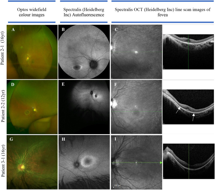
FIGURE 3.
Color fundus, autofluorescence, infrared, and OCT images in three individuals of two families with SRD5A3-CDG. (A) Optos image of eye with nystagmus (leading to some distortion of the image), demonstrating mild vascular attenuation, myopic oval disc morphology, and subtle macula reflex abnormalities. The peripheral retina does not reveal any pigmentary abnormalities; (B) delineating “watershed” zone between relatively preserved central retina, and dystrophic periphery; (C) loss of ellipsoid outside the perifoveal region in both eyes; (D) myopic discs, retinal nerve fiber layer reflexes constrained to macula, subtle vascular attenuation, and no significant retinal pigmentation migration suggestive of retinal dystrophy. (E) Abnormal macula with hyper-autofluorescent ring around fovea; (F) loss of ellipsoid—the outer retinal layers beyond the fovea (as highlighted by arrows). (G) Color image of left eye; (H) hyper-autofluorescent ring illustrating watershed area between preserved central macula function and dystrophic retinal periphery; (I) loss of ellipsoid layer outside the perifoveal retina.

