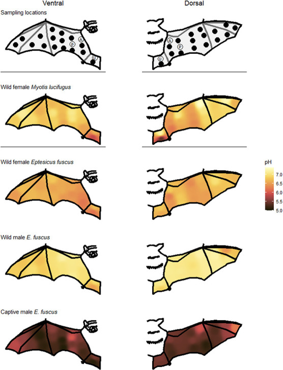
Figure 2.

Schematic views of the right wing and tail membrane indicating where we measured skin pH. All 38 measurements were taken from four individual bats while ‘P’ (ventral and dorsal plagiopatagium), ‘A’ (ventral and dorsal arm) and ‘U’ (ventral and dorsal uropatagium) were taken from all bats. Heat maps illustrate skin pH measurements taken from the ventral (left; 19 skin sites per bat) and dorsal (right; 19 skin sites per bat) flight membranes of bats caught in Ontario 2019. Myotis lucifugus and captive E. fuscus were measured in June and the two wild E. fuscus were measured in May.
