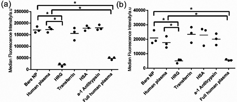
Figure 4.
Uptake of single protein corona-coated nanoparticles in liver (a) and brain endothelium (b). The 200 nm SiO2–NH2 were coated with 15 μg mL–1 human plasma, HRG, transferrin, HSA, or alpha-1 antitrypsin as described in the Materials and Methods section, and 30 μg mL–1 corona-coated nanoparticles were added to cells for 4 h in serum-free medium. As additional controls, the uptake in serum-free medium of 30 μg mL–1 bare nanoparticles (bare NP) and nanoparticles coated with full human plasma corona prepared as described in the Materials and Methods section (full human plasma) was also measured. The results of three independent experiments are shown, together with their average indicated with a line. A Kruskal–Wallis test was used to compare the different groups and indicated significant differences in both panels. A Mann–Whitney test with Bonferroni correction for multiple testing was applied to compare the uptake level of single protein corona-coated nanoparticles to the uptake of bare nanoparticles (bare NP) or nanoparticles coated with 15 μg mL–1 human plasma (human plasma). p ≤ 0.05 was considered significant (indicated with an asterisk).

