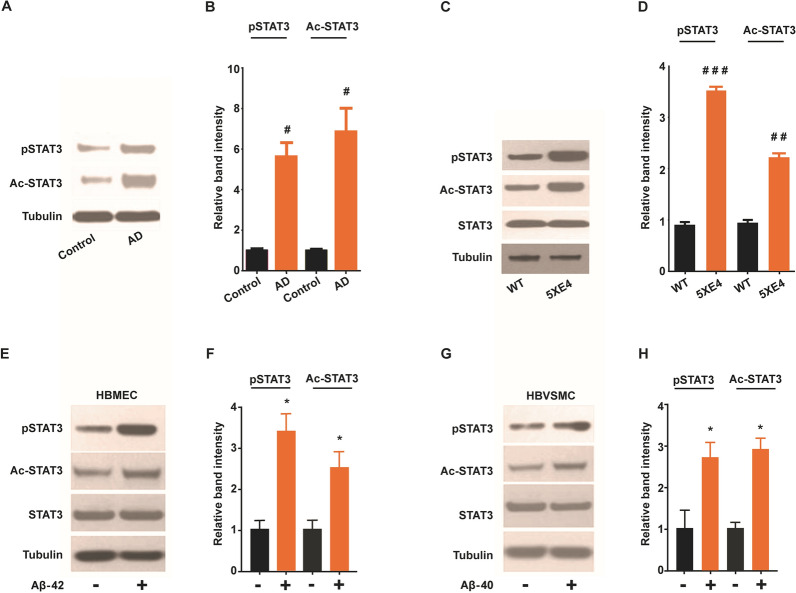
Fig. 1.
Activated forms of STAT3 is upregulated due to AD-related pathology. A, B Western blot analysis and quantification of STAT3 expression in blood vessels isolated from brain autopsy samples from age matched control and AD patients. C, D Immunoblot and quantification of STAT3 expression in blood vessels isolated from brains of 5XE4 and age matched wild-type (WT) mice. E, F Western blot analysis and quantification of STAT3 expression in cell homogenates following Aβ (1 µM) exposure to human brain microvascular endothelial (HBMEC). G, H Western blot analysis and quantification of STAT3 expression in cell homogenates following Aβ (1 µM) exposure to human brain vascular smooth muscle cells (HBVSMC). STAT3 phosphorylation at Tyr705 (pSTAT3); STAT3 acetylation at Lys685 (Ac-STAT3). Data is presented as mean ± SEM. #p < 0.05, ##p < 0.01, ###p < 0.001 as compared to control or WT, n = 4–5; *p < 0.05 as compared to no Aβ exposure. The cell culture experiments were performed in triplicate manner

