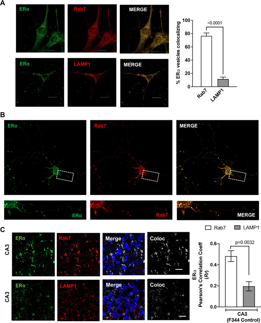
Figure 4.
ERα is localized on endolysosomes. A, Confocal images show the codistribution of ERα (green) with Rab7 or LAMP1 in CLU199 cells. Scale bar, 10 µm. Bar graph shows the quantification of object-based colocalization by reconstructing ERα, Rab7, and LAMP1 vesicles as spots in Imaris. B, In primary mice hippocampal neurons (DIV 9–11), ERα (green) codistributes with Rab7 (red). Scale bar, 10 µm. C, Confocal images show the colocalization of ERα (green) with Rab7 or LAMP1 in CA3 hippocampal region of F344 rat (male). Scale bar, 20 µm. Bar graph shows the quantification of colocalization by Pearson's correlation coefficient. At least 30 images were analyzed with 10–15 nuclei per image from five rats per group.

