
Figure 6.
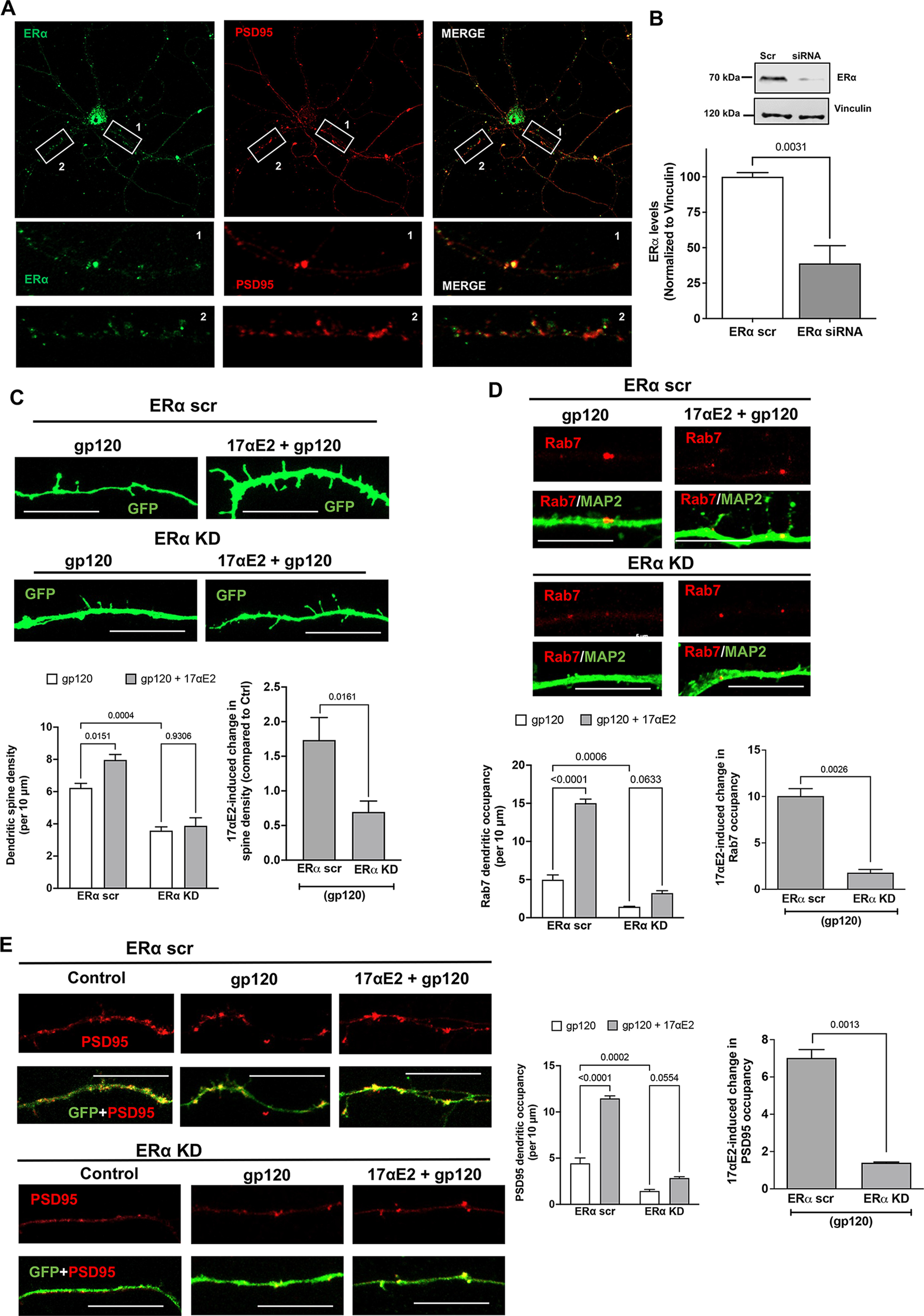
ERα knockdown prevents the protective effect of 17αE2 against HIV-1 gp120-induced impairment in dendritic spines. A, Immunostaining of ERα (green) and PSD-95 (red) in primary mouse hippocampal neurons (DIV 9–11). Colocalization of ERα and PSD-95 (75.16 ± 5.338%) along the dendrites is shown in enlarged panels. B, Densitometric quantification of immunoblotting show ERα protein levels in primary mouse hippocampal neurons (DIV 18) treated with ERα siRNA or scr siRNA; vinculin is used as a loading control (n = 4). C, Confocal images and bar graphs show that 17αE2 pretreatment (10 nm, 10 min) affects dendritic spine density (indicated by GFP expression) in ERα scr and ERα KD neurons (DIV 12–14) treated with HIV-1 gp120 (0.5 nm, 30 min). Bar graph on the right shows that ERα KD significantly reduced 17αE2-induced changes in spine density in gp120-treated neurons. D, Confocal images and bar graphs show that 17αE2 pretreatment (10 nm, 10 min) affects the occupancy of Rab-7-positive endolysosome in dendrites (identified with MAP2 staining) in ERα scr and ERα KD neurons (DIV 12–14) treated with HIV-1 gp120 (0.5 nm, 30 min). Bar graph on the right shows that ERα KD significantly reduced 17αE2-induced changes in the occupancy of Rab-7-positive endolysosome in dendrites in gp120-treated neurons. E, Confocal images and bar graphs show that 17αE2 pretreatment (10 nm, 10 min) affects the occupancy of PSD-95 (red) along dendrites (GFP) in both ERα scr and ERα KD neurons (DIV 12–14) treated with HIV-1 gp120 (0.5 nm, 30 min). Bar graph on the right shows that ERα KD significantly reduced 17αE2-induced changes in the occupancy of PSD-95 in dendrites in gp120-treated neurons.