
FIGURE 2.
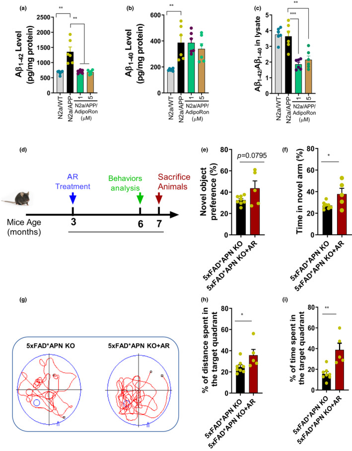
AR treatment reduced Aβ deposition and cognitive impairment. (a) The level of Aβ1‐42 in the cell lysate. (b) The level of Aβ1‐40 in the cell lysate. (c) The ratio of Aβ1‐42/Aβ1‐40. (d) 3‐month‐old 5xFAD*APN KO mice were orally treated with AR for 4 months. (e) The preference of the new object in the new object recognition test. (f) The percentage of time spent in a novel arm in the Y‐maze test. (g) The representative swimming trace in probe trial of morris water maze test. (h) The percentage of distance traveled in the target quadrant. (i) The percentage of time spent in the target quadrant. Data were expressed as mean ± SEM, * p < 0.05, ** p < 0.01, *** p < 0.001, **** p < 0.0001