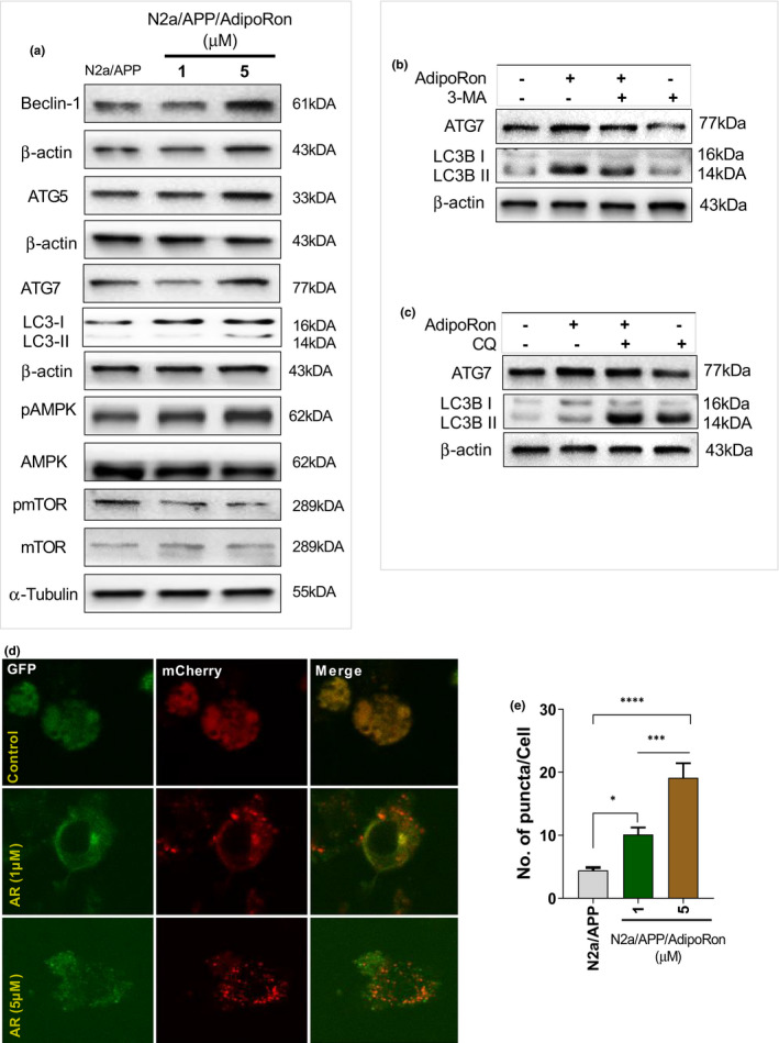
FIGURE 4.

AR significantly triggered autophagy by mediating AMPK‐mTOR pathway signaling. The level of Beclin‐1, ATG5, ATG7, LC3II/LC3I, pAMPK/AMPK, and pmTOR/mTOR in N2aAPPswe cells with or without AR treatment. (b) Autophagy inhibitor 3‐MA was used to suppress autophagy, representative Western blot analyses of LC3II and ATG7 levels. (c) Representative Western blot analyses of LC3II and ATG7 levels in the presence of CQ. (d‐e) Autophagosome images obtained with laser confocal microscopy after the N2aAPPswe cells were transfected with a mCherry‐GFP‐LC3B plasmid and treated with AR for 48 hours. Data were expressed as mean ± SEM, * p < 0.05, *** p < 0.001, **** p < 0.0001
