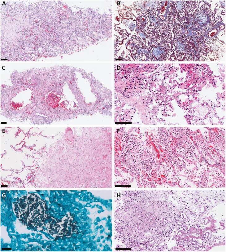
Figure 4.
Pulmonary histological findings of 80 fatal cases of coronavirus disease 2019 (COVID-19), ultrasound-guided minimally invasive tissue sampling. All scale bars = 100 µm unless otherwise specified. A, Severe proliferative/organizing diffuse alveolar damage with lymphocytic septal infiltrate, and fibroblast intra-alveolar proliferation, depositing collagen matrix. Hematoxylin and eosin (H&E) staining. B, Collagen matrix deposition in alveolar spaces. Masson trichrome staining. C, Severe interstitial fibrosis with “pseudo-cystic” formations due to barotrauma. H&E staining. D, Septal capillaries with angiomatoid aspect associated with tiny fibrin clots. H&E staining. E, Thrombus in a pulmonary artery, with surrounding parenchyma exhibiting coagulative necrosis. H&E staining. F, Secondary suppurative bacterial pneumonia. H&E staining. G, Grocott silver stain showing hyphae compatible with Candida spp in a case of COVID-19 with bacterial pneumonia. Scale bar = 20 µm. H, Pulmonary miliary tuberculosis associated with COVID-19. H&E staining.

