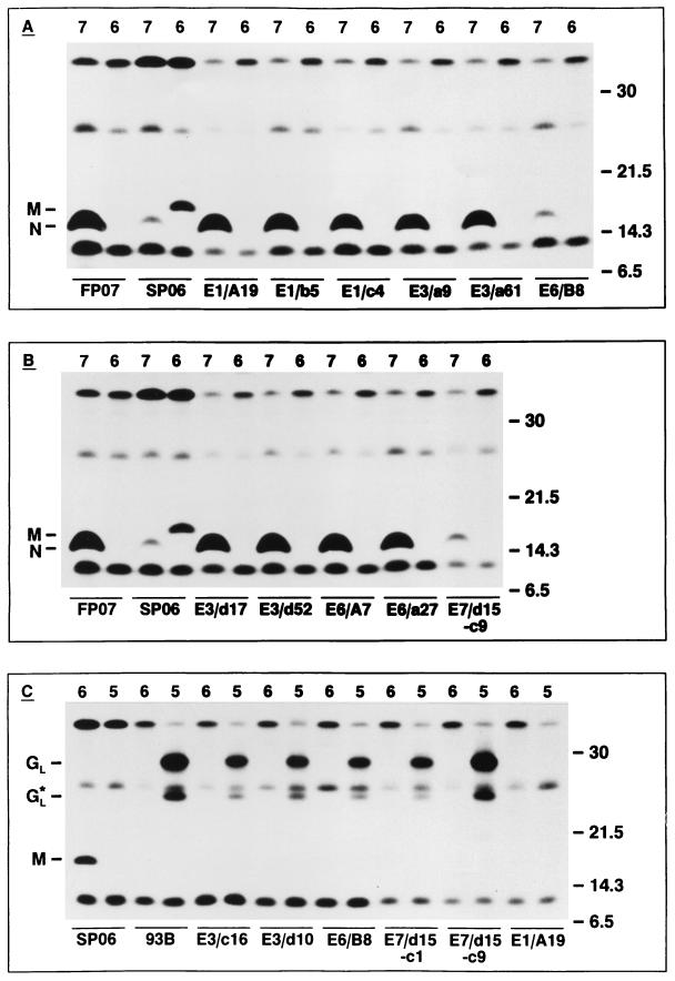
FIG. 2.
(A and B) Immunoprecipitation analysis of N-specific MAbs. OST-7.1 cells were infected with the vaccinia virus recombinants vAVE07 or vAVE16, which express the EAV N and M proteins, respectively, and were labeled for 1 h starting at 5 h p.i. with [35S]Met plus [35S]Cys. Total lysates of cells and media were prepared and incubated with 0.5 to 3 μl of N-specific (FP07) or M-specific (SP06) rabbit antiserum or 100 μl of culture supernatant from mouse hybridoma E1/A19, E1/b5, E1/c4, E3/a9, E3/a61, E3/d17, E3/d52, E6/A7, E6/a27, E6/B8, or E7/d15-c9. (C) Immunoprecipitation analysis of GL-specific MAbs. OST-7.1 cells were infected with the vaccinia virus recombinants vAVE16 (lanes 6) and vAVE25 (lanes 5), which express the EAV M and GL proteins, respectively (12), and were labeled as described above. Total lysates of cells and media were prepared and incubated with 0.5 μl of anti-M rabbit serum (SP06) or ascitic fluid containing the GL-specific MAb 93B (IgG2a subtype) (23) or 100 μl of culture supernatant from mouse hybridoma E3/c16, E3/d10, E6/B8, E7/d15-c1, E7/d15-c9, or E1/A19. The numbers at the right indicate the sizes (in kilodaltons) and positions of marker proteins analyzed in the same SDS–15% PAA gels. The positions of the N and M proteins and the full-length and a truncated form (GL*) (12) of the GL glycoprotein are displayed at the left.

