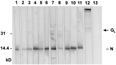
FIG. 3.
Immunoblot analysis of EAV-specific MAbs. Nitrocellulose strips containing electrophoretically separated virion proteins of EAV were incubated with MAbs E1/b5 (lane 1), E1/c4 (lane 2), E1/A19 (lane 3), E3/d17 (lane 4), E3/d52 (lane 5), E3/a61 (lane 6), E3/a9 (lane 7), E6/a27 (lane 8), E6/A7 (lane 9), and E6/d24 (lane 10, diluted 1:3; lane 11, diluted 1:8), with a mixture of MAbs E3/c16, E3/d10, and E6/B8 (lane 12), and with the anti-PRRSV MAb P11/d72 (lane 13) (50). The positions of the (putative) N and GL proteins of EAV are shown at the right side; the numbers at the left side indicate positions and sizes (in kilodaltons) of marker proteins analyzed in the same gel.

