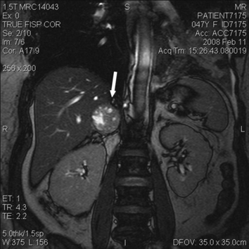
Figure 2.

Coronal view of the abdomen on magnetic resonance imaging with gadolinium contrast. The arrow points toward the right adrenal mass, which measures 4×3.7×4.2 cm; its proximity to the liver is evident. The lesion is predominantly solid but also contains multiple foci of high T2 signal intensity. The left adrenal gland is normal.
