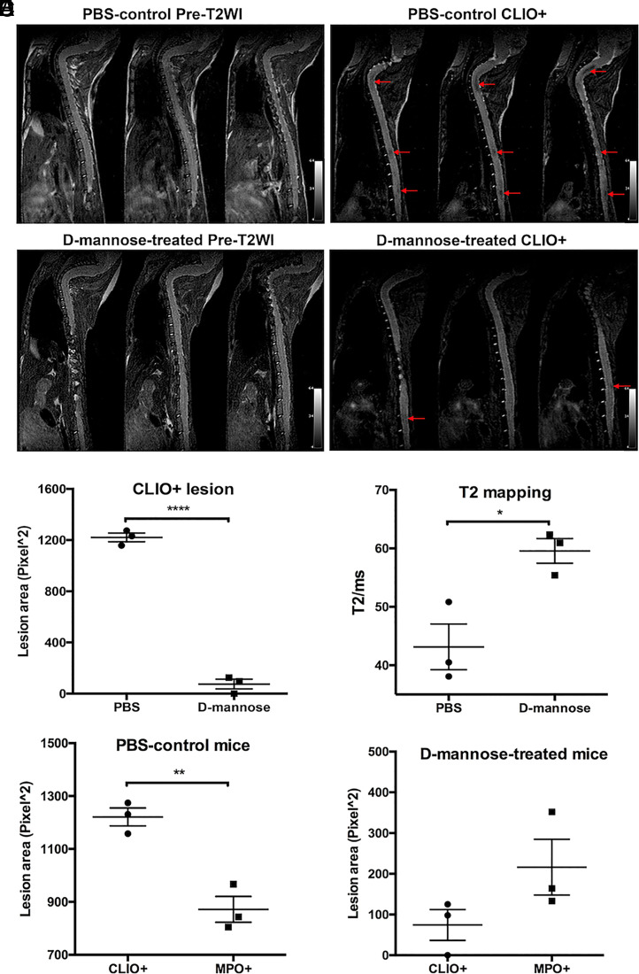
Fig. 3.
Phagocytosis imaging and analyses with CLIO nanoparticles. Representative consecutive slices of pre- and postcontrast enhanced T2-weighted MR imaging of (A) PBS-control and (B) d-mannose–treated mice. Low signal CLIO+ lesions (arrows) were found on postcontrast enhanced T2-weighted imaging in PBS-control mice and d-mannose–treated mice. (C) A significantly smaller total lesion area was found for d-mannose–treated mice than for PBS-control mice. (D) T2 values of the CLIO+ lesions in the PBS-control group of mice were significantly lower than those in the d-mannose–treated group of mice. (E) In PBS-control mice, the mean CLIO+ lesion area was larger than the mean MPO+ lesion area, while (F) in d-mannose–treated mice, the mean CLIO+ lesion area was smaller than the mean MPO+ lesion area. *P < 0.05, **P < 0.01, ****P < 0.0001.

