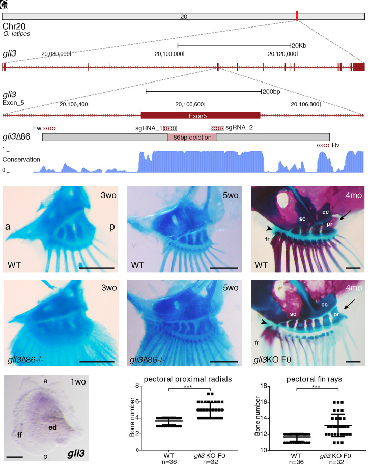
Fig. 1.
Medaka gli3 mutants show an increased number of pectoral fin skeletal elements. (A) Stable lines harboring Δ86 deletions in medaka gli3 exon 5 were generated by CRISPR-Cas9. The diagram shows the position of the deletion relative to the sgRNAs and primers used for screening along with the box of conservation with four fish species (stickleback, fugu, tetraodon, and zebrafish). The Δ86 deletion results in the generation of a premature STOP codon that truncates the predicted protein upstream of the zinc-finger DNA-binding domain of Gli3. (B–G) Alcian blue and Alizarin red staining of pectoral fin skeletons reveals a significantly increased number of proximal radial bones and fin rays in stable (E and F) and transient (G) gli3 mutants, although major anatomical AP asymmetries appear unaltered. Note that, when compared with ones in the anterior margin, the two posterior proximal radials (pr) are larger in both gli3 crispants and WT fish and articulate with the coracoid (cc) bone (arrows in D and G). In the anterior margin of the fin, both the rest of the proximal radials and the anterior-most fin rays (fr) articulate directly with the scapula (sc) in both WT and transient gli3 mutants (arrowheads in D and G). (B and C) n ≥ 14 fins. (E and F) n ≥ 12 fins. (Scale bars, 250 μm.) mo, months-old; wo, weeks-old. (H) WT pectoral fin showing the expression of gli3 in the anterior region of the developing pectoral fin bud (n = 10). (Scale bar, 100 μm.) ed, endochondral disk; ff, fin fold. (I and J) Quantification of skeletal elements in adult (4-mo-old) gli3 crispants. Each point in the graphs represents the measurement of bone number in a single fin (n = 36 WT, n = 32 gli3 KO F0). An unpaired t test was used for the statistical analysis of skeletal element number. ***P = 5.06 × 10⁻¹⁰ for the comparison between WT (mean 3.639) and gli3 crispant (mean 4.969) pectoral proximal radial bones. ***P = 2.33 × 10⁻⁷ for the comparison between WT (mean 11.67) and gli3 crispant (mean 13.13) pectoral fin rays. Bone-staining procedures in juvenile (B, C, E, and F) and adult fish (D and G) were performed in three independent experiments. a, anterior; p, posterior.

