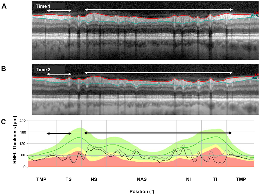
Figure 4.

An example of progression. (A) The circumpapillary b-scan from time 1. (B) The b-scan for time 2. C. The cRNFL thickness profiles for the first (gray) and second (black) times. The black and white arrows show regions of progression. (Eye ID 3)
