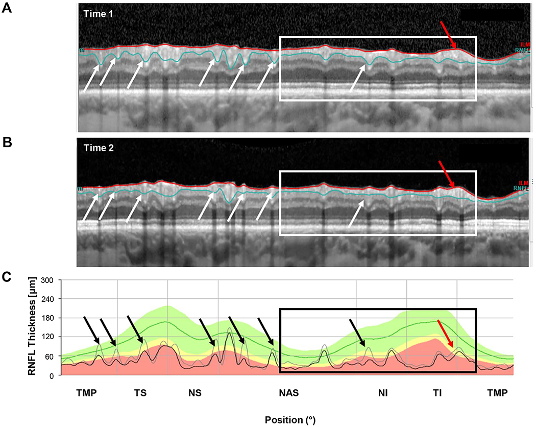
Figure 5.

An example of segmentation errors. (A) The circumpapillary b-scan from time 1. (B) The b-scan for time 2. C. The cRNFL thickness profiles for the first (gray) and second (black) times. The black and white arrows show regions where there are segmentation errors (time 1 includes less of the blood vessels than time 2). Progression can also be seen in the region within the black and white rectangles., especially where indicated by the red arrow. (Eye ID 2)
