Fig. 2.
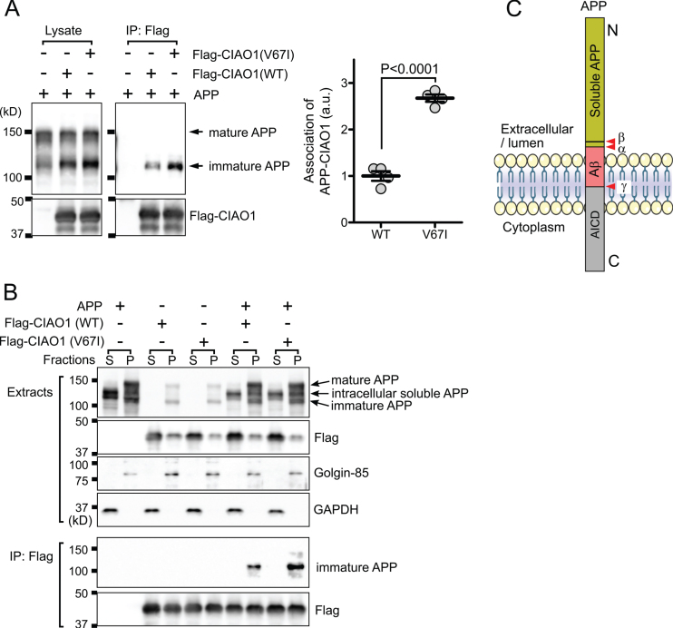
CIAO1 interacts with AβPP. A) Triton-soluble extracts of cells transfected with the indicated plasmids were immunoprecipitated using anti-flag magnetic beads (IP: Flag). Each precipitate was subjected to SDS-PAGE and western blotting. Arrows indicate the mature and immature forms of AβPP. The association of immature AβPP and CIAO1 was quantitatively analyzed in four independent experiments using Image J software. The association of AβPP with CIAO1-WT and CIAO1-V67I was statistically significant according to Student’s t-test. Arbitrary unit (a.u.). B) Cells transfected with the indicated plasmids were fractionated into cytosol (S) and membrane (P) fractions. Each form of AβPP is indicated by an arrow. Golgin-85 and GAPDH were used as marker proteins for the membrane and cytosol fractions, respectively. CIAO1 and the mutant were immunoprecipitated using anti-flag magnetic beads. C) Diagram of the AβPP structure. The cleavage sites for α-, β-, and γ-secretases are indicated (red arrowheads). AβPP intracellular domain (AICD). The anti-AβPP antibody used in this study was raised using a synthetic peptide corresponding to the juxtamembrane region surrounding at proline 620 of the human AβPP protein.

