
Fig. 3.
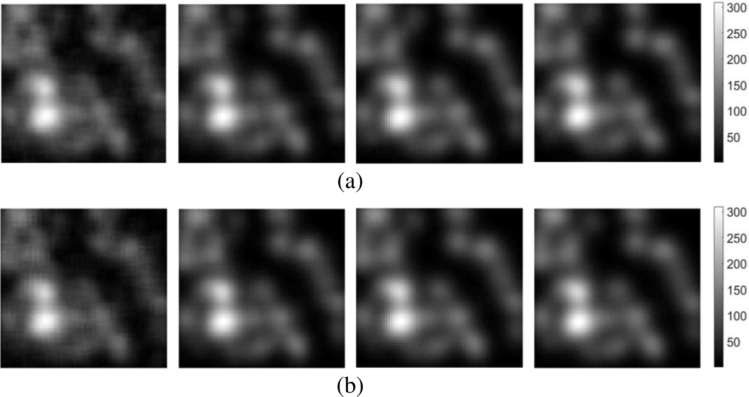
The images, from left to right, in each row represent the denoised estimates obtained by use of a) the CNN-based and b) the ResNet-based non-linear networks with varied {3, 7, 11, 13} layers, respectively. The related noise-free signal-present target image and the original noisy image g were the second and third images shown in Fig. 1. The dimensions of the images are 64 × 64.