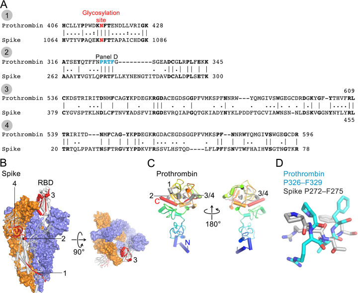
Fig 4. Sequence analysis of SARS-CoV-2 Spike and human prothrombin.
A. Sequence alignments of SARS-CoV-2 Spike (numbering corresponds to UniProt ID P0DTC2) and human prothrombin (P00734). B. Cryo-EM structure of trimeric Spike (Protein Data Bank ID 6Z97 [36]). Two protomers are shown in surface representation (blue and orange) and one as a grey cartoon with 4 peptide regions shown in red and indicated. Region 3 is located in the receptor-binding domain (RBD). Glycans are not shown for clarity. C. Crystal structure of prothrombin (PDB ID 5EDM, [35]) shown as a cartoon (N-terminus, blue; C-terminus, red). 4 peptide regions are shown in grey and indicated. D. Structural superposition of the PRTF motif from Spike and prothrombin.

