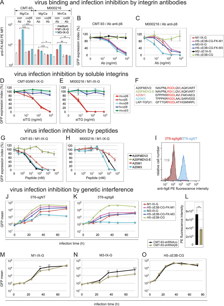
Fig 6. Inhibition of virus binding and infection by β6- and β8-specific antibodies, sITGs, peptides and genetic interference.
(A) Virus binding interference in CMT-93 and M000216 cells by β6/-β8 function blocking antibodies. Detached cells were sequentially incubated for 1 h on ice with control antibody, or the anti-β6/-β8 antibodies, followed by incubation with control medium or medium containing the indicated viruses at an MOI of 4, the rabbit anti-FK-M3 antibodies, and finally the secondary PE-conjugate antibodies. Incubation and washing buffer contained either Mg2+/Ca2+, 1 mM each, or 1/0.2 mM Mn2+/Ca2+. (B-C) Virus infection interference in CMT-93 and M000216 cells by β6- and β8-specific antibodies. CMT-93 (B) and M000216 cells (C) were pre-incubated for 1 h on ice using 5-fold dilution series of the specific β6- or β8-integrin antibodies, respectively, starting with 800 ng/ml as highest concentration, followed by addition of the different indicated GFP-expressing viruses and transfer to 37°C for 48 h. An MOI of 1 was used for CMT-93 cells and MOI of 3 for M000216 cells in all experiments shown in this figure. GFP analysis was performed 48 h pi, and expression index was normalized to a control antibody. IC50 values determined in this experiment are summarized in Table 2. (D, E) Infection blocking assays by sITGs. M1-IX-G virus was incubated for 1 h at RT with 5-fold serial dilutions of the indicated sITGs starting from 800 ng/ml to 6.4 ng/ml, followed by addition to CMT-93 cells (D) and M00216 cells (E) and cultivated and further processed as above. (F-H) Infection blocking assays by peptides. (F) The 20-mer peptides tested for virus infection inhibition included peptides A20FMDV2 derived from the VP1 coat protein of FMDV2, A20FMDV2-E containing a D to E mutation in the critical RGD motif, A20M1 and A20M3 derived from M1-/M3-FK, respectively, as compared to LAP-hTGFβ1, all containing the critical αvβ6/αvβ8-binding RGDLXX(L/I) motif. (G, H) Cells were pre-incubated on ice with 5-fold serial dilutions of peptides resulting in final concentrations from 5,000 to 0.32 nM. Subsequently, M1-IX-G virus was added to CMT-93 cells (G) or M000216 cells (H), followed by processing as described above. (I) Comparative flow cytometry profiles of αvβ8 expression in 3T6 cells. Blue and red show β8-specific staining in 3T6-sgNT and 3T6-sgItgβ8 cells, respectively, and grey histogram shows background staining of 3T6-sgItgβ8 cells using a matched isotype control. Numbers indicate MFI values of specific or control antibodies. (J, K) Transduction of 3T6-sgNT and 3T6-sgItgβ8 cells using M1-/M3-IX-G, the fiber-chimeric H5-ΔE3B-CG-FK-M1/-FK-M3 and control H5-ΔE3B-CG at an MOI of 9. Cells were processed as described in Fig 1B. (L) Comparative flow cytometry MFI αvβ6 expression values in control CMT-93-sgNT versus β6 integrin shRNA knock down CMT-93-sgItgβ6 cells. (M-O) Infection of control CMT-93-sgNT and CMT-93-sgItgβ6 cells using M1-IX-G (M), M3-IX-G (N) and H5-ΔE3B-CG (O) at an MOI of 1. Cells were processed as described above. Except for the representative flow cytometry histogram in (I), data represent triplicates, shown as mean ± SEM. Asterisks indicate level of significance for comparison of indicated values (*, P<0.05; **, P<0.005; ***, P<0.0005).

