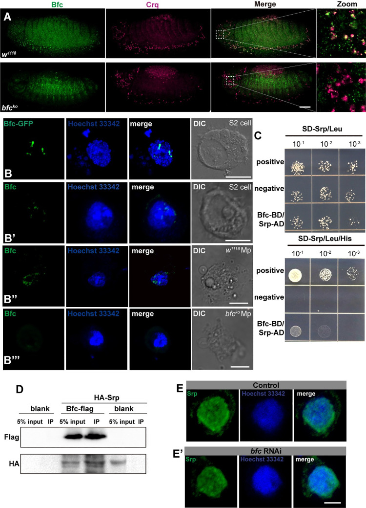
Fig 4. bfc is expressed in embryonic macrophages, and physically interacts with the GATA factor Srp.
A. Macrophages of stage 13 w1118 and bfcko embryos were stained with anti-Crq (magenta) and anti-Bfc (green) antibodies. Scale bar = 20 μm. B. Representative fluorescence pictures of the Bfc-GFP expression in S2 cells; B’-B”‘. Anti-Bfc (green) stained S2 cells (B’); macrophages (showed Mp for short in figure) isolated from w1118 larvae (B”) and bfcko larvae (B”‘); the nuclei were stained using Hoechst 33342 (blue). Scale bar = 5 μm. C. Yeast two-hybrid assays to detect the interaction of Bfc (Bait) with Srp (Prey). Different concentrations of the labeled yeast transformants were grown on SD-Trp-Leu-His plates. D. Crude protein extracts from blank S2 cells, transiently transfected S2 cells expressing Bfc-Flag, and HA-Srp or HA-Srp alone were immunoprecipitated with anti-Flag magnetic beads. WB detection was performed using anti-Flag and anti-HA antibodies. E-E’. Anti-Srp (green) stained control S2 cells as well as bfc RNAi-treated S2 cells; the nuclei were stained using Hoechst 33342 (blue). Scale bar = 5 μm.

