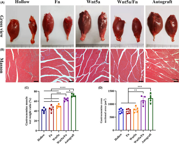
FIGURE 7.

Gastrocnemius muscle recovery 12 weeks after the operation. (A) Representative gross views of gastrocnemius muscles of each group. The injured side was on the right. The normal side was on the left. (B) Representative images of Masson staining in each group. Original magnification is 100 ×. Scale bar =100 μm. (C) The wet weight ratio of the gastrocnemius (injured side/the normal side) of each group. (D) The mean cross‐sectional area of muscle fibers of each group. *p < 0.05, **p < 0.01, ***p < 0.001. Data are expressed as the mean ±SD. Fn, fibrin hydrogel group
