
FIGURE 6.
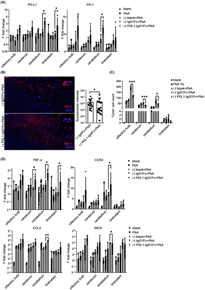
Intracerebral enhancement of PD‐L1 affected the immune microenvironment of each brain region. Intrathecal injection was performed at −1 dpi and 1 dpi on C57BL/6 mice. (A) The mRNA levels of PD‐L1 are upregulated in the cerebellum and brainstem while PD‐1 increased in the cerebellum of ECM mice injected with PDL1‐IgG1Fc, with the IgG1Fc group as control. IF‐stained brain sections at 7 dpi indicate increased PD‐1 (B) and decreased TUNEL (C) staining in the PDL1‐IgG1Fc group compared with that in the IgG1Fc group. (D) The changes in mRNA levels of inflammatory cytokines are shown in the brain of ECM mice injected with PDL1‐IgG1Fc compared with those in the IgG1Fc group. The results are expressed as the mean ± SD of three independent experiments. *p < 0.05, **p < 0.01, and ***p < 0.001 indicate that the differences are significant (unpaired t‐test, n = 3)