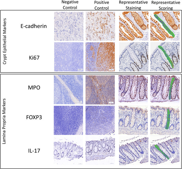
Figure 2.

Representative immunohistochemical staining for rectal crypt epithelium markers E‐cadherin and Ki67 and stromal markers MPO, FOXP3 and IL‐17 within human colorectal mucosa. Tissue is from MSM engaging in CRAI. Positive control tissues are breast cancer tissue for E‐cadherin, tonsillar tissue for Ki67, MPO and FOXP3 and upper GI tissue for IL‐17 as described in the Methods section. Negative control staining was done with non‐immune IgG as described in the Methods section. All histologic sections were counterstained with haematoxylin. Images also depict aspects of image analyses to measure the optical densities of the biomarkers in colonic hemicrypts (epithelial markers) and in the areas adjacent to the hemicrypts (lamina propria markers) outlined in green and separated into 50 equidistant segments from the base of the crypt area to the lumen. A minimum of three regions were scored per biomarker, per participant visit.
