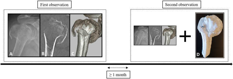
Fig. 1.
During the first observation, proximal humerus fractures were assessed with conventional onscreen imaging. During the second observation, 3D-printed models were added. The image labeled with the letter A represents the trauma radiograph, B the 2D CT image (coronal plane), C the 3D CT image (anterolateral aspect), and D the 3D-printed handheld model. A color image accompanies the online version of this article.

