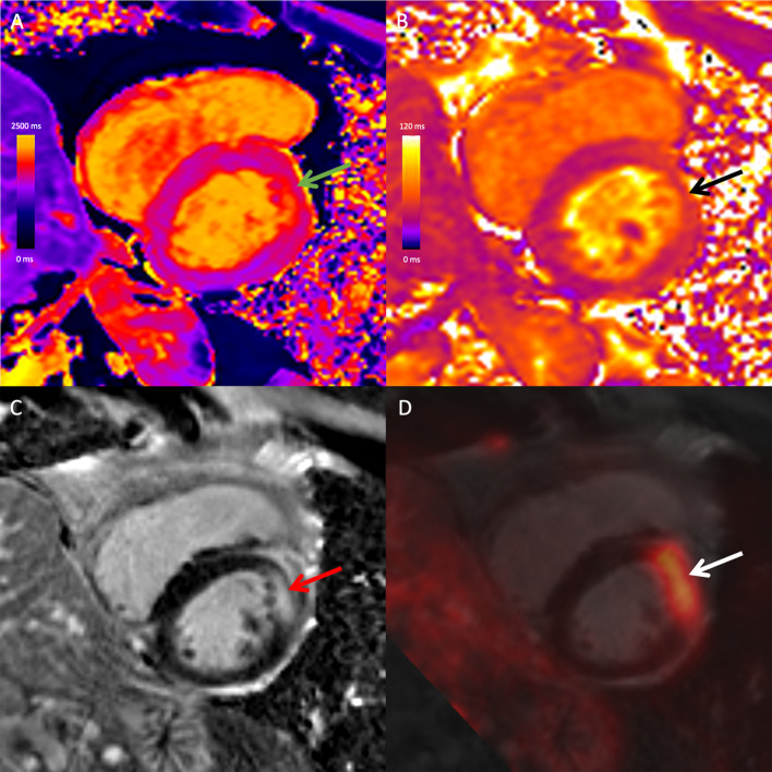
Fig. 1.
Combined 18F-FDG PET/MRI images in a 67-year-old male with cardiac and extra-cardiac sarcoidosis. Mid-ventricular short-axis native T1 map (A), native T2 map (B), late gadolinium-enhanced (LGE) image (C), and fused 18F-FDG PET and LGE image (D) demonstrate high T1 (green arrow), high T2 (black arrow), mid-wall LGE (red arrow) and co-localizing focal FDG uptake (white arrow) at the mid-anterior wall. There was extra-cardiac FDG uptake in mediastinal and hilar lymph nodes (not shown). The study was positive on both PET and MRI, likely reflecting active cardiac sarcoidosis with myocardial inflammation

