Fig. 6.
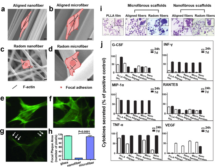
The effects of hydrogel architecture on cell activities. a–d Schematic image of the interaction between cell and hydrogel scaffolds with different topographies. Cells generally exhibited a spindle-shaped morphology on microfibers (b, d) or aligned fibers (a, b), while evolved into the rounded morphology on nanofibers or randomly oriented fibers (c). Reproduced with permission.249,419 Copyright 2013, Wiley-VCH. The figure is made with biorender (https://biorender.com/). e–h Fibroblast F-actin staining (green) on glass (e), microfiber (f), and nanofiber (g), as well as the quantification analysis of focal plaque area (h). The arrows represent membrane protrusions (“cork-screw” ruffles). Reproduced with permission.30 Copyright 2011, Wolters Kluwer Health, Inc. i, j Evidences on how the architecture of hydrogel scaffolds regulated macrophage morphology (i) and cytokine expression (j), where microfibers (1–50 μm) induced M1 microphage activations and more proinflammatory cytokine secretions compared to fibers with a diameter of 200–600 nm. Adapted with permission.257 Copyright 2011, American Chemical Society

