Figure 5.
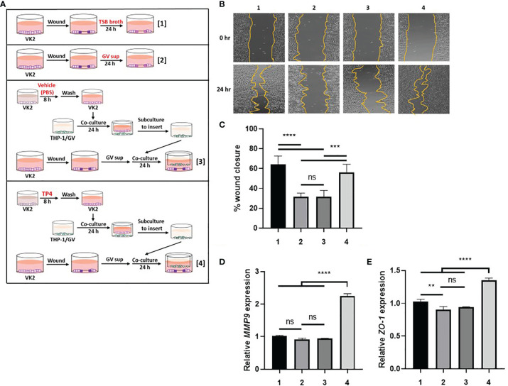
TP4-induced M2a macrophages promote VK2 cell migration and epithelial barrier integrity. (A) Schematic description of the treatment methods. VK2 cell monolayers were scratched with a sterile pipette and then treated with [treatment 1] TSB broth or [treatment 2] 10% (v/v) GV sup. Meanwhile, GV sup-treated VK2 cells were co-cultured with THP-1/GV cells which had been incubated with [treatment 3] vehicle (PBS)- or [treatment 4] TP4 (7.82 µg/ml)-treated VK2 cells. (B) Wound-healing assays were performed at 0 and 24 h in VK2 cells. (C) Wound closure was calculated as the percent area remaining uncovered by the cells at the given timepoint. (D) MMP9 expression was measured by qRT-PCR after 24 h co-culture. (E) ZO-1 expression was measured by qRT-PCR after 48 h co-culture. Data were represented the normalized target gene amount relative to the group of treatment 1. Data are presented as mean ± SD of three independent experiments (**P < 0.01; ***P < 0.001; ****P < 0.0001; ns, not statistically significant).

