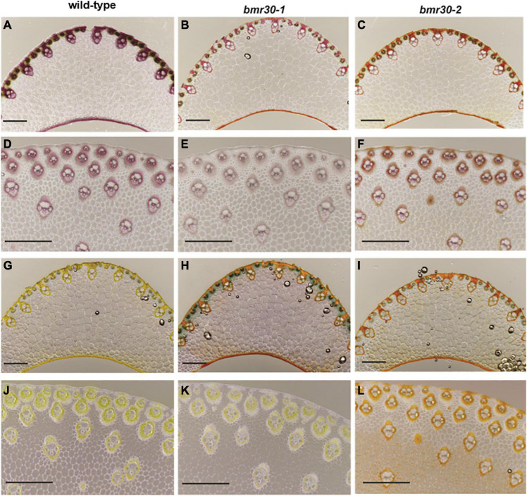
FIGURE 4.
Visualization of lignification using phloroglucinol (A–F) and vanillin-HCl (G–L) staining of leaf midrib (A–C,G–I) and stalk (D–F,J–L) tissues from wild-type (BTx623), bmr30-1, and bmr30-2 plants. Scale bar = 500 μm for both leaf midrib and stalk tissue. Leaf midrib tissue was observed at 40× magnification, stalk tissue was observed at 80× magnification using an Olympus BX-51 light microscope (Olympus Co.).

