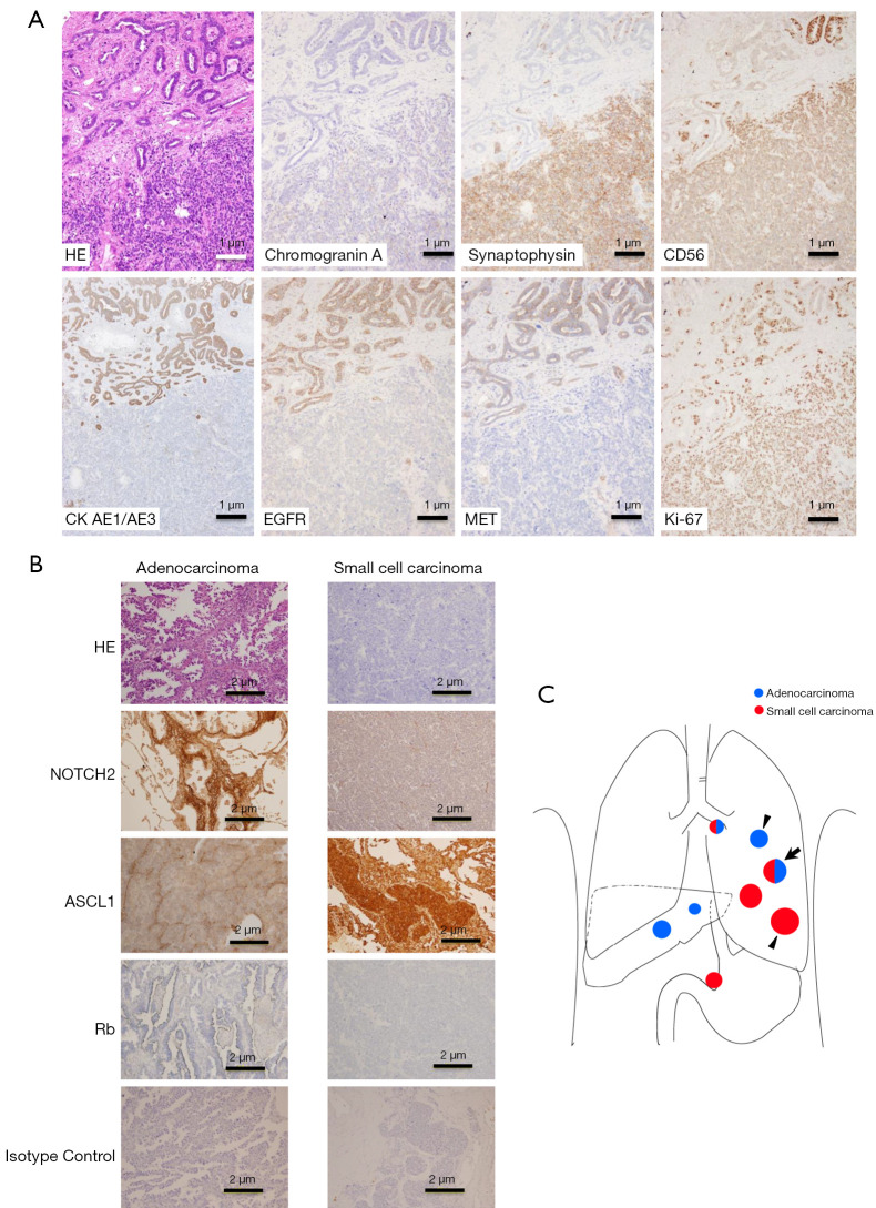
Figure 3.
Autopsy findings in case 1. (A) Immunohistochemical staining at the border of coexistent metastasis in the left upper lung lobe, indicated as arrow in (C). Adenocarcinoma in the upper region is in contact with the small-cell carcinoma in the lower region. (B) Immunohistochemical staining of isolated histological lesions indicated as arrowheads in (C). (C) Schema of tumor distribution at case 1 autopsy. HE, hematoxylin-eosin; CK, cytokeratin; EGFR, epidermal growth factor receptor; MET, mesenchymal-to-epithelial transition; ASCL1, Achaete-scute homolog 1; Rb, retinoblastoma.

